- Christina Sumners
- Biosciences & Technology, Research, Show on VR homepage
Improving chemotherapy health outcomes
Texas A&M researchers show why targeting a gene called RASSF1A can reduce the deleterious effects of malignant tumors during chemotherapy
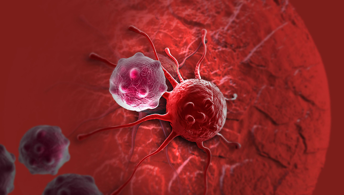
cancer cell
Chemotherapy is an effective way to kill rapidly dividing cells in malignant tumors. But a major side effect of chemotherapeutic drugs is that they cause inflammation, which then damages normal, healthy cells. To solve this hurdle in cancer treatment, scientists at the Center for Translational Cancer Research at the Texas A&M Institute of Biosciences and Technology have identified a biotarget that can reduce inflammation during chemotherapy.
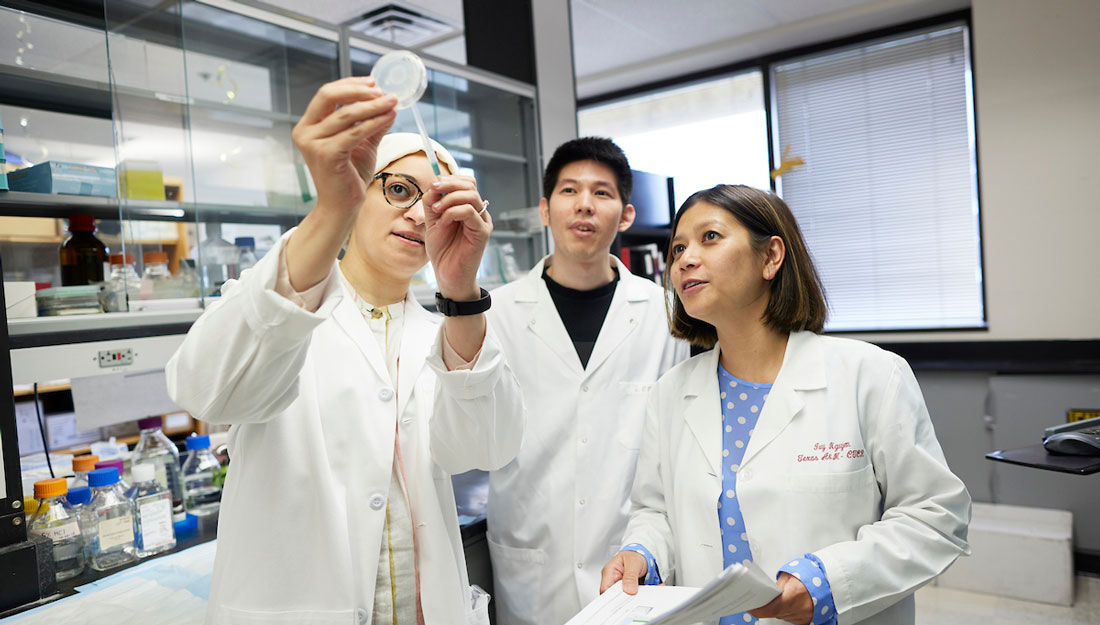
Using a variety of genetic tools, including CRISPR-Cas, the Texas A&M team investigated the cellular mechanisms by which a tumor suppressing gene called RASSF1A operates in cancer cells. In their article, published in the journal Cell Death & Differentiation, they show that RASSF1A reduces cancer malignancy by activating autophagy—a cellular process that plays a key role in regulating inflammation.
Autophagy is a mechanism by which cells “reduce, reuse and recycle” their biological content. Structures known as autophagosomes transport defunct cellular materials (DNA, RNA, proteins, lipids, etc.) to organelles called lysosomes for degradation. Since autophagy serves necessary housekeeping functions in cells, cancer research has largely focused on developing autophagy-inhibiting drugs to kill malignant tumors.
However, suppressing autophagy can have unwanted secondary effects. When cancer cells die, they send distress signals to the immune system eliciting inflammation.
Since cancer cells have diminished autophagy compared to normal cells, researchers are now looking at ways to improve autophagy. “Our current understanding is that we need cancer cells to have an active autophagy,” said Leyuan Liu, PhD, assistant professor and senior author of the study. “We need to enhance autophagy in such a way that cancer cells become less malignant.”
Earlier work from Liu’s laboratory showed that a protein called MAP1S activates autophagy. MAP1S interacts with many other autophageal proteins, including the protein coded by the RASSF1A gene. This observation led his team to investigate if the cellular mechanisms by which RASSF1A regulates autophagy is similar to MAP1S.
Liu’s team found that RASSF1A coordinates autophagy through a different cellular process. When RASSF1A was absent in cells, the streamline flow of autophagosomes to the lysosome is severely disrupted. “Without RASSF1A, autophagosomes stay where they are,” Liu said. “In other words, they cannot get into the lysosome for degradation.”
They also observed that cellular deficits in RASSF1A and, by extension, autophagy led to the rampant growth of liver cancer in animal models.
Unlike many tumor suppressive genes, including MAP1S, RASSF1A is not mutated in cancer cells. The levels of RASSF1A are just lower in malignant tumors. Liu noted that restoring RASSF1A to normal levels may thus hold the key to reducing malignancy.
“If you can develop a drug that can activate RASSF1A and restore autophagy function, then that is new course for cancer treatment,” he said.
Liu also said that both RASSF1A and MAP1S could be targeted independently since the two coordinate autophagy through different cellular mechanisms.
Liu hopes that autophagy-facilitating treatments will be able to reduce the adverse effects of chemotherapy in patients. “Killing cancer cells is relatively easy. But what happens to the host after killing cancer cells is more important,” Liu explained. “Our goal is to reduce the bad impact of cancer cells on the host. In my opinion, we need to think about the host in addition to ways to deal with cancer cells.”
Article written by Vandana Suresh
Media contact: media@tamu.edu